Omentum
The greater omentum is a sheet of mesentery covered by peritoneal epithelium and filled with fatty, loose connective tissue. It hangs from the lower border of the transverse colon, forming a fatty apron-like structure within the abdomen. This is where fat is stored, which can contribute to the increased abdominal girth seen in obese middle-aged individuals.
Vascularization
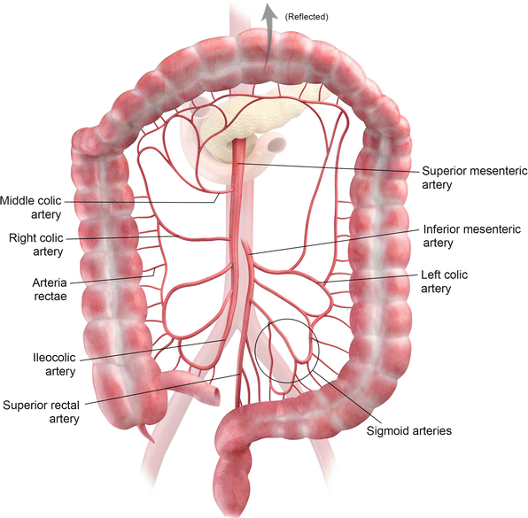
The blood supply to the colon is provided by the superior mesenteric artery (SMA) and the inferior mesenteric artery (IMA). These two vessels are connected via the marginal artery, which runs alongside the entire length of the colon. The specific branches supplying different parts of the bowel are as follows:
- The cecum receives blood from the ileocolic artery, a terminal branch of the SMA. The ileocolic artery also gives rise to the appendicular artery, which supplies the appendix.
- The ascending colon and right colic flexure are supplied by the ileocolic and right colic arteries, both of which originate from the SMA.
- The arterial supply to the transverse colon primarily comes from the middle colic artery, a branch of the SMA. It may also receive blood from the anastomotic arcades formed by the right and left colic arteries, which together create the marginal artery.
- The descending and sigmoid colon are supplied by the left colic and sigmoid arteries, both branches of the IMA. The change in blood supply at the left colic flexure, where it transitions from the SMA to the IMA, marks the embryological shift from the midgut to the hindgut.
- The rectum and anal canal receive blood from the superior rectal artery, a continuation of the IMA, along with branches from the internal iliac arteries—the middle and inferior rectal arteries. The inferior rectal artery is a branch of the internal pudendal artery.
Watershed areas: Two main areas in the colon, including splenic flexure (Griffiths point) and rectosigmoid junction (Sudek’s point), are prone to ischemia. The reason is that both are the transition points from 2 major supplying arteries–> Splenic flexure is the area between SMA and IMA supplies, and the rectosigmoid junction is the region between the IMA and the superior rectal artery supplies. These areas mostly supplied by the marginal artery; however, in 50% of the population, this artery is poorly developed.
Venous drainage follows a pattern similar to arterial supply. The inferior mesenteric vein (IMV) drains into the splenic vein, while the superior mesenteric vein (SMV) joins the splenic vein to form the hepatic portal vein. Lymphatic drainage of the large intestine flows into the lymph nodes associated with these primary vessels.
Rectum peculiarity: The rectum has two drainage veins. The upper and middle thirds of the rectum drain primarily into the superior rectal vein and finally empty into the liver via the inferior mesenteric vein and portal vein. However, the lower third of the rectum drains into the middle rectal vein. The blood in the middle rectal vein skips the liver because it drains directly into the IVC. This is the reason that suppository medications are rapidily absorved, bypass the liver following systemic absorption, which reduces the hepatic first-pass effect.
Nerves
The midgut-derived structures, including the ascending colon and the proximal two-thirds of the transverse colon, receive their parasympathetic, sympathetic, and sensory innervation from the superior mesenteric plexus.
The hindgut-derived structures, including the distal one-third of the transverse colon, the descending colon, and the sigmoid colon, receive parasympathetic, sympathetic, and sensory innervation from the inferior mesenteric plexus.
